Latvian artificial intelligence solution developer "Apply" in cooperation with radiologists - assoc. Prof. Kārlis Kupčs and Dr. Andris Veiss - have created the solution "Synapse", which with the help of computer vision will help health care specialists in hospitals to diagnose strokes faster. Since May 2024, the solution has been used at Paula Stradiņš Clinical University Hospital, currently showing 80% efficiency.
Like the human brain, artificial intelligence also makes a decision as a result of sequential analysis of facts. "Synapse" consists of several neural networks and combinations of several computer vision algorithms, each of which has a specific task. Each layer of the brain is analyzed in an attempt to understand whether pathology might be present there. Since each person's brain is different, it is important for the solution to learn to adapt to their specifics.
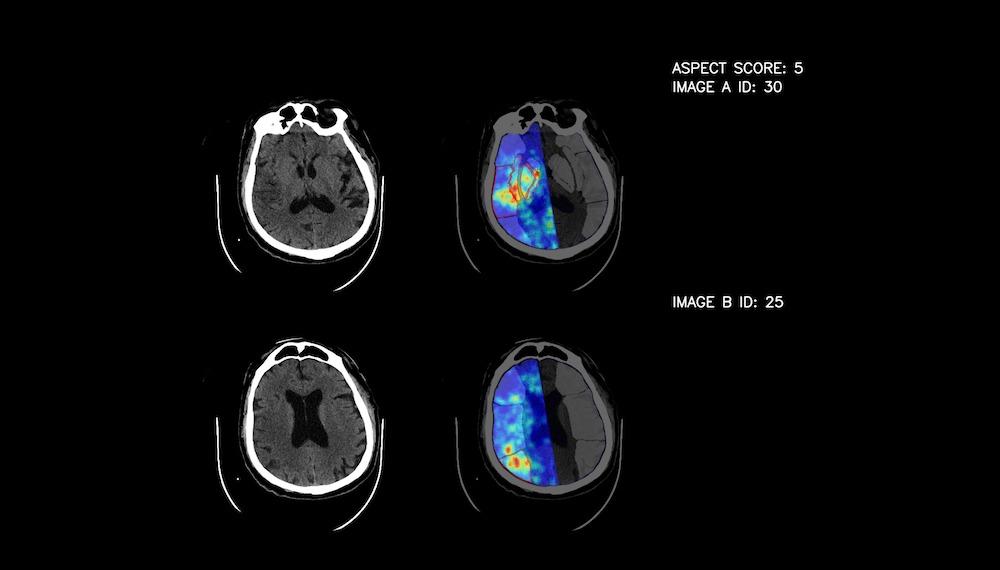
When analyzing examinations, "Synapse" also helps to calculate the "aspect number" used internationally in radiology, marking the seriousness of the patient's condition. As the information is collected, the algorithm evaluates all the available information, determining whether the patient might have had a stroke and which areas of the brain were affected. Through interactions between clinicians and AI solution developers, the “training” or calibration process continues, improving the solution's analytics capabilities.
The beginnings of the project can be traced back to 2020, thanks to the support of the Competence Center for Pharmaceutical, Biomedical and Medical Technologies. Until now, the solution developed by "Apply" provides the highest accuracy among global competitors, with the prototype of the solution successfully recognizing 80% of stroke cases. According to the radiologists, the most successful competitor's solution, which the hospital managed to try so far, showed only a 60% success rate.
Improves diagnostic accuracy
Radiologist, Assoc. prof. Kārlis Kupčs says that a stroke, or blockage of cerebral arteries, can be treated very successfully these days, the main thing is to go to the hospital in time. After a stroke, approximately two million human nerve cells die every minute. Although there are billions of such cells in our brain, each of them is responsible for its own function.
"During the first hours of an ischemic stroke, changes in computed tomography examinations are practically invisible or very minimal, and it is challenging to recognize them with the naked eye. It requires the experience of knowledgeable specialists and the analysis of complex symptoms," he said. The new solution is designed to help radiologists by improving diagnostic accuracy. This will draw attention to whether an ischemic stroke is potentially observed in the specific computed tomography examination, while the doctor's task is to confirm this assumption. Computed tomography examination evaluation takes approximately three minutes for the solution.
"The solution will not only reduce the diagnostic time, but will be especially useful at times when the doctor's attention is slowed down, even during the 24-hour duty. It is also hoped that in the near future this solution will be useful for regional hospitals, where the shortage of personnel, including doctors, is more pronounced, and the radiologist often interprets examinations remotely," says K. Kupčs.
Ischemic stroke is the second most common cause of death and the third most common cause of disability in the world, including in Latvia. Unfortunately, it can leave irreversible consequences in the body - speech and movement disorders, paralysis of the body on one or both sides of the body, etc. To prevent these risks, it is important to get medical help in time. The best results are achieved if the treatment is started within the first two hours after the stroke.
Helps doctors in their work
In the case of a stroke, the patient's journey consists of various complex stages and specialists. Since "speed" is the key word in the treatment process, the diagnostic time saved with the help of artificial intelligence can significantly shorten the total time until the patient receives help.
K. Kupčs believes that "Synapse" is an excellent addition to the "sight" of an experienced radiologist. The solution proved its importance already in the first times when the solution was used for examination analysis. "Currently, we have signed an agreement with Paula Stradiņš Clinical University Hospital for further testing of the solution prototype. By integrating the tool into the hospital's existing system, it helps our doctors' work on a daily basis," he says.
The solution is to keep learning
"Apply" project manager Līna Briņže emphasizes that no solution and no equipment will be able to replace the doctor. However, AI can be as sharp as a doctor, so its main goal is to become a doctor's assistant and helper.
“It is quite challenging to teach the solution to 'see' the way healthcare professionals do. Medical data is very specific and complex, therefore we constantly cooperate with radiologists during the project development stage to ensure the most accurate operation of the solution," said L. Briņģe.
"Apply's" experience in the development of various types of computer vision solutions, including health care projects, allowed us to reach a successful solution relatively quickly. At the moment, more than 400 data samples have already been processed, and thanks to the opportunity to test the prototype of the solution at the Paula Stradiņš Clinical University Hospital, it is planned to allow the system to learn the same number of data samples.
"It should be noted that the learning process is never-ending and data uploads will continue throughout the 'lifetime' of the solution. The more data, the more accurate and widely applicable the solution will become," adds L.Briņģe
It is expected that by developing this solution, in the future "Apply" will be able to develop an equivalent method for the diagnosis of other examinations.
Photo: publicity photo